A) saddle
B) hinge
C) pivot
D) plane
E) ball and socket
G) B) and C)
Correct Answer

verified
Correct Answer
verified
Multiple Choice
The type of movement between carpal bones is described as
A) pivot.
B) adduction.
C) extension.
D) flexion.
E) gliding.
G) B) and C)
Correct Answer

verified
Correct Answer
verified
Multiple Choice
Standing on one's toes is an example of a movement called
A) dorsiflexion.
B) plantar flexion.
C) depression.
D) opposition.
E) elevation.
G) B) and C)
Correct Answer

verified
Correct Answer
verified
Multiple Choice
Returning the thumb to the anatomical position after touching the little finger is
A) reposition.
B) opposition.
C) medial excursion.
D) supination.
F) A) and D)
Correct Answer

verified
Correct Answer
verified
Multiple Choice
Cartilaginous joints
A) are common in the skull.
B) unite two bones by means of fibrocartilage or hyaline cartilage.
C) allow the most movement between bones.
D) are found in the lower leg.
E) are not found in the pelvic region.
G) A) and B)
Correct Answer

verified
Correct Answer
verified
Multiple Choice
When two bones grow together across a joint to form a single bone, this is called a
A) suture.
B) syndesmosis.
C) gomphosis.
D) synostosis.
E) symphysis.
G) B) and C)
Correct Answer

verified
Correct Answer
verified
Multiple Choice
A tennis player goes to the doctor and is told he has a torn rotator cuff. He has injured his
A) neck.
B) shoulder.
C) hip.
D) knee.
E) elbow.
G) A) and C)
Correct Answer

verified
Correct Answer
verified
Multiple Choice
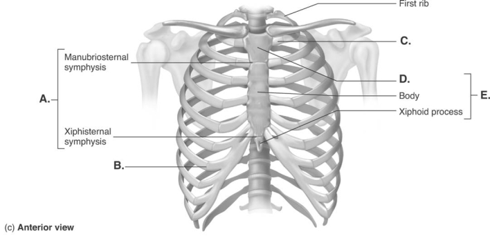
 -The figure illustrates the joints and bones of the rib cage. What does "E" represent?
A) costochondral joint
B) sternum
C) manubrium
D) sternal symphyses
E) sternocostal synchrondrosis
G) None of the above
Correct Answer

verified
Correct Answer
verified
Multiple Choice
 -The figure illustrates the joints and bones of the rib cage. What does "A" represent?
A) costochondral joint
B) sternum
C) manubrium
D) sternal symphyses
E) sternocostal synchrondrosis
G) B) and E)
Correct Answer

verified
Correct Answer
verified
Multiple Choice
An example of a saddle joint is the
A) shoulder joint.
B) elbow joint.
C) atlanto-occipital joint.
D) carpometacarpal joint.
E) atlantoaxial joint.
G) B) and E)
Correct Answer

verified
Correct Answer
verified
Multiple Choice
A pivot joint
A) is a modified ball and socket joint.
B) restricts movement to rotation.
C) is a biaxial joint.
D) allows gliding movement.
E) is between the atlas and the occipital bone.
G) C) and D)
Correct Answer

verified
Correct Answer
verified
Multiple Choice
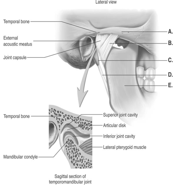
 -The figure illustrates structures in the right temporomandibular joint (lateral view) . What does "D" represent?
A) lateral ligament
B) mandible
C) zygomatic arch
D) styloid process
E) stylomandibular ligament
G) A) and E)
Correct Answer

verified
Correct Answer
verified
Multiple Choice
Articular cartilage
A) is a double layer of tissue that encloses a joint.
B) is a thin lubricating film covering the surface of a joint.
C) provides a smooth surface where bones meet.
D) is a layer of tissue that is continuous with the periosteum.
E) lines the joint everywhere except over the articular cartilage.
G) A) and B)
Correct Answer

verified
Correct Answer
verified
Multiple Choice
A movement through 360 degrees that combines flexion, extension, abduction, and adduction is called
A) circumduction.
B) rotation.
C) hyperextension.
D) supination.
E) pronation.
G) A) and E)
Correct Answer

verified
Correct Answer
verified
Multiple Choice
Moving the mandible to the side as when grinding the teeth is
A) lateral flexion.
B) lateral excursion.
C) elevation.
D) inversion.
F) A) and C)
Correct Answer

verified
Correct Answer
verified
Multiple Choice
Which of the following statements regarding the temporomandibular (TMJ) joint is correct?
A) The joint is divided into lateral and medial cavities by an articular disc of cartilage.
B) The joint has a cartilage capsule.
C) The joint is a combination plane and ellipsoidal joint.
D) The joint allows rotation.
E) The joint is located between the maxilla and the mandible.
G) A) and D)
Correct Answer

verified
Correct Answer
verified
Multiple Choice
Osteoarthritis is
A) a bacterial infection transmitted by ticks.
B) an inflammation of any joint.
C) a metabolic disorder caused by increased uric acid in blood.
D) a condition that may involve an autoimmune disease.
E) the most common type of arthritis.
G) B) and E)
Correct Answer

verified
Correct Answer
verified
Multiple Choice
Gout is
A) a bacterial infection transmitted by ticks.
B) an inflammation of any joint.
C) a metabolic disorder caused by increased uric acid in blood.
D) a condition that may involve an autoimmune disease.
E) the most common type of arthritis.
G) None of the above
Correct Answer

verified
Correct Answer
verified
Multiple Choice
 -The figure illustrates structures in the right temporomandibular joint (lateral view) . What does "B" represent?
A) lateral ligament
B) mandible
C) zygomatic arch
D) styloid process
E) stylomandibular ligament
G) B) and C)
Correct Answer

verified
Correct Answer
verified
Multiple Choice
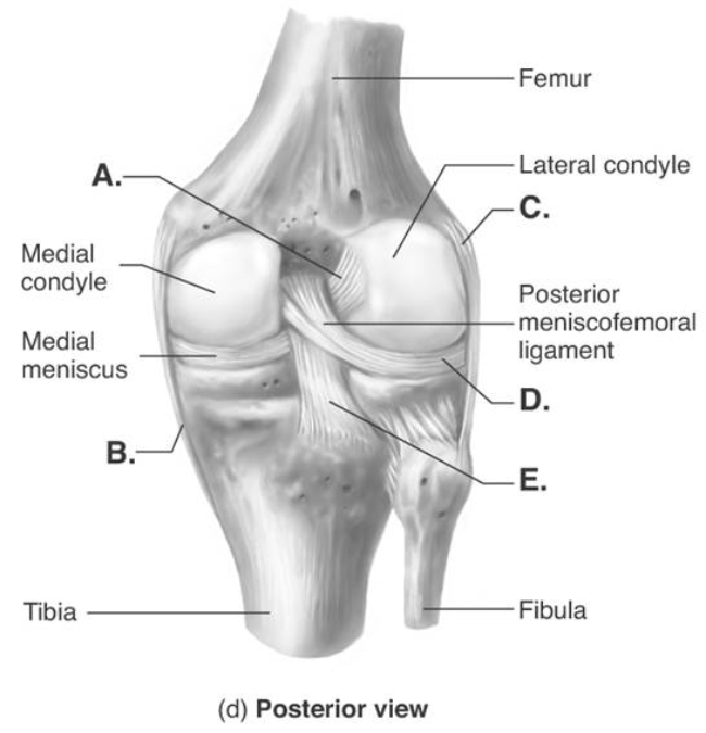
 -The figure illustrates a posterior view of the right knee joint. What does "C" represent?
A) medial (tibial) collateral ligament (MCL)
B) posterior cruciate ligament (PCL)
C) anterior cruciate ligament (ACL)
D) lateral (fibular) collateral ligament (LCL)
E) lateral meniscus
G) A) and E)
Correct Answer

verified
Correct Answer
verified
Showing 21 - 40 of 119
Related Exams